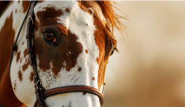
The first thing anyone notices is the size. Horse eyes are large — obviously, strikingly large compared to nearly any other land mammal. But horse eye anatomy goes well beyond raw size. Every structure, from the horizontally oriented pupil to the reflective layer behind the retina, is a precisely tuned evolutionary response to the demands of life as a large prey animal in open terrain.
Understanding horse eye anatomy isn’t anatomy for its own sake. It’s the foundation for understanding how horses perceive the world, why they behave the way they do, and what to watch for when something changes in how they see.
How big are horse eyes compared to other animals?
At approximately 5 centimeters (2 inches) in diameter, the horse’s eye is the largest of any land mammal. For context:
- Human eye: approximately 2.5 cm
- Dog eye (medium breed): approximately 1.8 to 2.0 cm
- Elephant eye: approximately 3.7 cm — surprisingly smaller than the horse’s, despite the massive body size difference
This scale isn’t cosmetic. A larger eye gathers more photons per unit of time, which directly improves performance in low-light conditions. A larger pupil at maximum dilation means more incoming light during twilight and pre-dawn — exactly the hours when many predators are most active.
Why does the horse have a horizontal pupil?
The horse’s pupil is a horizontal oval — an elongated slit running transversely across the iris. This shape is shared by most grazing prey mammals (sheep, goats, deer, antelope) and stands in sharp contrast to the circular pupils of humans and the vertical slits of many predators.
The horizontal pupil serves two functions simultaneously:
Maximizing lateral light intake: the wide horizontal opening captures more light from the sides of the visual field, amplifying the sensitivity of the monocular lateral zone — exactly where a prey animal needs maximum motion detection.
Natural glare control: the horizontal format acts as a built-in visor, reducing excessive light entry from above (direct sunlight) and below (ground reflection) while maintaining a wide lateral aperture. This is critical for an animal that spends hours in open fields under direct sun.
Does the horse’s pupil stay horizontal when the head tilts?
Yes — and this is one of the most remarkable details of equine visual anatomy. Research has shown that when a horse tilts its head laterally, the muscles of the eye globe actively rotate the eye to maintain the pupil’s horizontal orientation relative to the ground.
This compensatory mechanism — ocular counter-rotation — ensures that the functional advantages of the horizontal pupil are preserved regardless of head position. When the horse lowers its head to graze or tilts it sideways to investigate something, the pupil stays horizontal automatically.
What makes the equine retina distinctive?
The retina is the photoreceptor layer at the back of the eye that converts light into neural signals. In horses, three features make it anatomically and functionally unusual:
High rod density: rod cells are the photoreceptors responsible for low-light vision. They don’t detect color, but they’re extremely sensitive to light variation. The equine retina has a much higher rod-to-cone ratio than the human retina, which is why horses navigate effectively in conditions that would leave humans visually impaired.
Dichromatic cone system: cones handle color processing and fine detail but require more light to function. Horses have only two types of cones — sensitive to short wavelengths (blue) and medium wavelengths (yellow-green) — making them dichromats. They perceive color, but with a narrower spectrum than humans, and cannot reliably distinguish red from green.
The visual streak: instead of a central fovea (the small fixed point of maximum acuity that anchors human vision), the equine retina has a horizontal band of elevated acuity running across the retina laterally. This band aligns with the horizon when the horse holds its head in a natural grazing position — maximum resolution precisely where a predator approach is most likely. It’s a distribution of visual resources calibrated to a specific threat environment.
What is the tapetum lucidum and how does it function?
The tapetum lucidum is a layer of reflective tissue positioned behind the retina. Normally, light entering the eye passes through the retina once — the photoreceptors capture what they can, and the rest is absorbed. The tapetum lucidum reflects that uncaptured light back through the retina a second time, giving photoreceptors another chance to register it. This effectively doubles the eye’s light-gathering efficiency in dim conditions.
In horses, the tapetum lucidum covers the upper half of the retina — the portion that receives light from below the horizon line (ground-reflected light, which is typically dimmer than overhead light). The lower half of the retina, which receives direct overhead and sky light, lacks the tapetum — avoiding amplification of already-abundant daytime illumination.
This asymmetrical arrangement is functionally elegant: it maximizes sensitivity exactly where light is weakest, without creating glare in conditions where light is plentiful.
The tapetum is also responsible for the characteristic eye shine horses produce when a direct light source hits them in the dark. The same structure creates eye shine in cats, dogs, and most nocturnal and crepuscular mammals.
What is the nictitating membrane?
Horses have a third eyelid — the nictitating membrane, or membrana nictitans — positioned at the medial corner of the eye, closest to the nose. This translucent structure can slide across the eye surface from the inner corner outward, covering the globe.
Functions of the nictitating membrane:
- Cleaning and lubricating the surface of the eye
- Mechanical protection against dust, debris, and vegetation
- Distributing tear film evenly across the cornea
In a healthy resting horse, the nictitating membrane is nearly invisible. When it becomes visible at rest — especially if it covers a significant portion of the iris — this is a potential indicator of eye irritation, infection, pain, or systemic illness, and warrants veterinary evaluation.
What eye conditions commonly affect horses?
The horse’s eye, by virtue of its size and lateral positioning, is more exposed to environmental trauma than the eyes of most other domestic species. Common conditions include:
Equine recurrent uveitis (ERU): also known historically as “moon blindness,” this is the most common cause of blindness in horses worldwide. Characterized by recurring bouts of intraocular inflammation, often associated with Leptospira bacteria. Progressive and vision-threatening without appropriate management.
Corneal ulcers: the exposed lateral eye position makes horses vulnerable to corneal trauma from hay, branches, dust particles, and insects. Ulcers that aren’t treated promptly can escalate to serious infection within days.
Cataracts: lens opacity, more common in older horses, that progressively reduces visual acuity. May manifest behaviorally as increased spooking in variable-light environments before vision loss becomes obvious to observers.
How good is horse visual acuity?
Equine visual acuity — the ability to resolve fine detail — is estimated at approximately 20/33 to 20/60 in human terms. This means a horse needs to be 20 feet from an object to see it with the clarity a person with normal vision would have at 33 to 60 feet.
In practice, horses see shapes, movement, and contrast clearly but don’t resolve fine detail at distance. A horse cannot read facial expressions across a field.
What it trades in acuity, however, it more than compensates for in other ways: the breadth of its visual field (nearly 350 degrees), its extreme sensitivity to motion at the periphery, and its capacity to function at light levels that would leave humans effectively blind.
This calibration makes biological sense. For a prey animal, detecting a predator early — when it’s still a silhouette at the edge of the field — matters far more than identifying it precisely once it’s already close. The system is built for early warning, not for identification.
Horse eye anatomy as a window into evolution
Every feature of the equine eye — the outsized globe, the horizontally rotating pupil, the visual-streak retina, the asymmetrically placed tapetum lucidum, the protective nictitating membrane — is a solution to a specific survival challenge. Horse eye anatomy is one of the most thoroughly studied among domestic mammals precisely because it reveals, in concrete structural detail, how evolutionary pressure shapes biological design.
For the equestrian professional or serious horse owner, this knowledge reframes behavior. When the horse responds the way it does — spooking at a flag, hesitating at a dark entrance, reacting differently to the same object on each side — it’s not random. It’s the output of a visual system that was refined, over millions of years, to do exactly what it does.